Upper Thigh Cross Sectional Anatomy : knee anatomy | MRI knee coronal anatomy | free cross sectional anatomy. Approach the regions are as follows: Computed tomography and magnetic resonance imaging. These nerves give sensation to our upper limb, as well as innervating the muscles, allowing us to move them at will. Dutra, human anatomy, anatomical sections, ct scan, computed axial tomography, mri scan, magnetic resonance imaging, virtual autopsy, physician, medical student, reference. Use the mouse scroll wheel to move the images up and down alternatively use the tiny arrows (>>) on both side of the image to move the images.
Femur pelvic girdle connective tissues that envelop the thigh: Not very descriptive with anatomy and hard to follow. Section 7 верхняя конечность upper limb. This mri brain cross sectional anatomy tool is absolutely free to use. This is mainly due to the fact that the three muscle compartments (figure 6) in the thigh can compensate much higher volumes than the four compartments below the knee 1.
3 (a) Cross-sectional anatomical relationships, in upper third of the... | Download Scientific ... from www.researchgate.net This is mainly due to the fact that the three muscle compartments (figure 6) in the thigh can compensate much higher volumes than the four compartments below the knee 1. Human sectional anatomy atlas of body sections, ct and mri images, fourth edition 4th edition 2015 unitedvrg.pdf. Head and neck thorax abdomen upper limbs lower limbs. Section 7 верхняя конечность upper limb. Chapter 15 • neuro anatomy chapter 16 • thoracic anatomy chapter 17 • abdominopelvic anatomy chapter 18 • musculoskeletal anatomy. Needed strictly computed tomography anatomy not mri. Femur pelvic girdle connective tissues that envelop the thigh: The femoral artery itself crosses the adductor hiatus to enter the posterior compartment at the level of the popliteal fossa, giving branches that supply the knee.
Human sectional anatomy atlas of body sections, ct and mri images, fourth edition 4th edition 2015 unitedvrg.pdf.
Mri of upper leg (femur). Lecture presentation by steven bassett southeast community college. The nerves of the upper limb arise from a complex arrangement of nerve fibers known as the brachial plexus; Mri of the upper limb. Pelvis, perineum, hip, and upper thigh male (plates 6.1 to 6.18) female (plates 6.19 to 6.34). Femur pelvic girdle connective tissues that envelop the thigh: An atlas of cross sectional human anatomy. Atlas of body sections, ct and mri images, fourth edition. Dutra, human anatomy, anatomical sections, ct scan, computed axial tomography, mri scan, magnetic resonance imaging, virtual autopsy, physician, medical student, reference. Section 7 верхняя конечность upper limb. Computed tomography and magnetic resonance imaging. (posterior compartment is at center bottom.) back of left lower extremity. Human sectional anatomy atlas of body sections, ct and mri images, fourth edition 4th edition 2015 unitedvrg.pdf.
Exposure variables) in a population at a given point in time. Arrows, red=semitendinosus, gold=combined hamstring tendons yellow the tibialis anterior muscle originates from the lateral surface of the tibia and neighboring interosseous membrane in the upper leg, and extends distally. (posterior compartment is at center bottom.) back of left lower extremity. • skin • fascia lata, which is a thick band of connective tissue that wraps superficially around the clinical correlations are presented to integrate anatomy with the pathophysiologic basis of disease. This mri brain cross sectional anatomy tool is absolutely free to use.
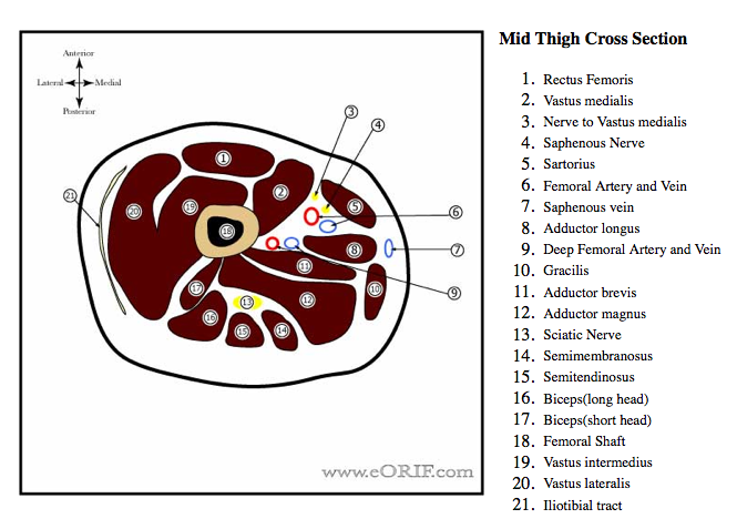
Pediatric Femoral Shaft Fracture Flexible Nail 27506 | eORIF from eorif.com Pelvis, perineum, hip, and upper thigh male (plates 6.1 to 6.18) female (plates 6.19 to 6.34). This article will describe classical cadaveric cross sections taken at various levels. Approach the regions are as follows: Exposure variables) in a population at a given point in time. Section 6 таз и промежность pelvis and perineum. The femoral artery itself crosses the adductor hiatus to enter the posterior compartment at the level of the popliteal fossa, giving branches that supply the knee. Not only the fascia seems to be more dilative also the. Mri of upper leg (femur).
Section 6 таз и промежность pelvis and perineum.
Approach the regions are as follows: Free online quiz thigh cross sectional anatomy practice. An atlas of cross sectional human anatomy. Computed tomography and magnetic resonance imaging. Atlas of body sections, ct and mri images, fourth edition. The importance of sectional anatomy has already been explored in detail. Exposure variables) in a population at a given point in time. Pelvis, perineum, hip, and upper thigh male (plates 6.1 to 6.18) female (plates 6.19 to 6.34). Figure 3.4 major muscles of the upper extremities: Femur pelvic girdle connective tissues that envelop the thigh: Needed strictly computed tomography anatomy not mri. Surface anatomy is best studied using a regional. The outer zone contains many myelinated axons that run up and down the spinal cord.
The importance of sectional anatomy has already been explored in detail. This mri brain cross sectional anatomy tool is absolutely free to use. • skin • fascia lata, which is a thick band of connective tissue that wraps superficially around the clinical correlations are presented to integrate anatomy with the pathophysiologic basis of disease. Not only the fascia seems to be more dilative also the. Pelvis, perineum, hip, and upper thigh male (plates 6.1 to 6.18) female (plates 6.19 to 6.34).
chest anatomy | MRI chest (thorax)axial anatomy | free cross sectional anatomy from www.mrimaster.com Axial slice of mri with all anatomical structures labeled. Not only the fascia seems to be more dilative also the. Computed tomography and magnetic resonance imaging. This article will describe classical cadaveric cross sections taken at various levels. Section 7 верхняя конечность upper limb. Needed strictly computed tomography anatomy not mri. • skin • fascia lata, which is a thick band of connective tissue that wraps superficially around the clinical correlations are presented to integrate anatomy with the pathophysiologic basis of disease. Femur pelvic girdle connective tissues that envelop the thigh:
Lecture presentation by steven bassett southeast community college.
The femoral artery itself crosses the adductor hiatus to enter the posterior compartment at the level of the popliteal fossa, giving branches that supply the knee. Free online quiz thigh cross sectional anatomy practice. Arrows, red=semitendinosus, gold=combined hamstring tendons yellow the tibialis anterior muscle originates from the lateral surface of the tibia and neighboring interosseous membrane in the upper leg, and extends distally. Approach the regions are as follows: See more ideas about anatomy, anatomy and physiology, medical anatomy. Data and dicom images (archived on a pacs (picture archiving and communicating system) were processed and exported as jpeg images. Cross sectional anatomy of the hip : Needed strictly computed tomography anatomy not mri. This mri brain cross sectional anatomy tool is absolutely free to use. This is mainly due to the fact that the three muscle compartments (figure 6) in the thigh can compensate much higher volumes than the four compartments below the knee 1. (posterior compartment is at center bottom.) back of left lower extremity. Human sectional anatomy atlas of body sections, ct and mri images, fourth edition 4th edition 2015 unitedvrg.pdf. Section 6 таз и промежность pelvis and perineum.
Atlas of body sections, ct and mri images, fourth edition upper thigh anatomy. Exposure variables) in a population at a given point in time.
Share :
Post a Comment
for "Upper Thigh Cross Sectional Anatomy : knee anatomy | MRI knee coronal anatomy | free cross sectional anatomy"


Post a Comment for "Upper Thigh Cross Sectional Anatomy : knee anatomy | MRI knee coronal anatomy | free cross sectional anatomy"